所員紹介
遠藤 斗志也(タンパク質動態研究所 所長)
研究内容
真核生物の細胞内には膜で仕切られたオルガネラ構造が発達している。オルガネラは固有のタンパク質群を備え、それらがオルガネラ固有の機能を実現すると共に、細胞内および細胞外環境からの要請に応答することで、細胞レベルの恒常性を維持している。オルガネラを構成するタンパク質が、どのようにオルガネラに移行し、オルガネラ内の適切な区画に仕分けられ、どのように機能構造を獲得し、複合体を形成するのか、これらのプロセスを担うタンパク質複合体の構造機能相関はどのようなものなのか,さらには核ゲノムとミトコンドリアDNAがコードするタンパク質が強調してつくられ働く仕組みはどのようなものかを解明しようとしている。
主な業績
- Endo T, Wiedemann N (2025)
Mechanisms and pathways of mitochondrial protein transport. Nat. Rev. Mol. Cell Biol. in press. - Takeda H, Busto JV, Lindau C, Tsutsumi A, Tomii K, Imai K, Yamamori Y, Hirokawa T, Motono C, Ganesan I, Wenz, L-S, Becker T, Kikkawa M, Pfanner N, Wiedemann N, Endo T (2023)
A multipoint guidance mechanism for β-barrel folding on the SAM complex. Nat Struct Mol Biol 30, 176-187. - H. Takeda, A. Tsutsumi, T. Nishizawa, C. Lindau, J.V. Busto, L.-S. Wenz, L. Ellenrieder, K. Imai, S.P. Straub, W. Mossmann, J. Qiu, Y. Yamamori, K. Tomii, J. Suzuki, T. Murata, S. Ogasawara, O. Nureki, T. Becker, N. Pfanner, N. Wiedemann, M. Kikkawa, and T. Endo (2021)
Mitochondrial sorting and assembly machinery operates by β-barrel switching. Nature 590 (7844), 163-169 - Y. Araiso, A. Tsutsumi, J. .Qiu, K. Imai, T. Shiota, J. Song, C. Lindau, L.-S. Wenz, H. Sakaue, K. Yunoki, S. Kawano, J. Suzuki, M. Wischnewski, C. Schütze, H. Ariyama, T. Ando, T. Becker, T. Lithgow, N. Wiedemann, N. Pfanner, M. Kikkawa, and T. Endo (2019)
Structure of the mitochondrial import gate reveas distinct preprotein paths. Nature 575, 395-401. - Sakaue H, Shiota T, Ishizaka N, Kawano S, Tamura Y, Tan KS, Imai K, Motono C, Hirokawa T, Taki K, Miyata N, Kuge O, Lithgow T, and Endo T. (2019) Porin associates with Tom22 to regulate the mitochondrial protein gate assembly. Mol Cell. 73, 1044-1055.
- Kawano S., Tamura Y., Kojima R., Bala S., Asai E., Michel A. H., Kornmann B., Riezman I., Riezman H., Sakae Y., Okamoto Y., and Endo T. (2018)
Structure-function insights into direct lipid transfer between membranes by Mmm1-Mdm12 of ERMES. J.Cell Biol. 217, 959-974
.
横山 謙 教授(生命科学部 先端生命科学科 教授)
研究内容
生命のエネルギー通貨であるATPは、主にミトコンドリアに存在するATP合成酵素により合成され、あらゆる生命現象のエネルギー源として使われる。一方、ATPは体内の色々な細胞や組織間の情報伝達に使われていることも知られている。このATPの意外な働きは、半世紀前から徐々に明らかにされてきたが、細胞内や細胞間でのATP動態の実態の解明は解決すべき問題として残されている。生命にとって最も重要な物質である ATPの動態をシステムと捉え(ATPシステム)、ATPシステムを担う膜タンパク質の構造を、クライオ電顕による構造解析で決定し、その構造的基盤を明らかにする。
主な業績
- Nakanishi A, Kishikawa J, Tamakoshi M, Mitsuoka K, & Yokoyama K. (2018)
Cryo EM structure of intact rotary H+-ATPase/synthase from Thermus thermophiles
Nat. Commun. 9, 02553-6 - Baba M, Iwamoto K, Iino R, Ueno H, Hara M, Nakanishi A, Kishikawa J, Noji H, & Yokoyama K. (2016)
Rotation of artificial rotor axles in rotary molecular motors
Proc. Natl. Acad. Sci. U S A 113, 11214-11219 - Furuike S, Nakano M, Adachi K, Noji H, Kinosita K. Jr, & Yokoyama K. (2011)
Resolving stepping rotation in Thermus thermophilus H+-ATPase/synthase with an essentially drag free probe
Nat. Commun. 2, 233-239 - Maher MJ, Akimoto S, Iwata M, Nagata K, Hori Y, Yoshida M, Yokoyama S, Iwata S, Yokoyama K. (2009)
Crystal structure of A3B3 complex of V-ATPase from Thermus thermophilus.
EMBO J. 28, 3771-3779 - Jormakka M, Yokoyama K., Yano T, Tamakoshi M, Akimoto S, Shimamura T, Curmi P, Iwata S. (2008)
Molecular mechanism of energy conservation in polysulfide respiration.
Nat. Struct. Mol. Biol.15:730-737. - Toei, H., Gerle, C., Nakano, N., Tani, K., Gyobu, N., Tamakoshi, M., Nobuhito Sone, N., Yoshida, M., Fujiyoshi, Y., Mitsuoka, K., and Yokoyama K. (2007) Dodecamer rotor ring defines H+/ATP ratio for ATP synthesis of prokaryotic V-ATPase from Thermus thermophilus.
Proc Natl Acad Sci U S A., Vol. 103, P 20256 –20261
津下 英明(生命科学部 先端生命科学科 教授)
研究内容
細菌は様々なタンパク毒素を用いて宿主細胞を攻撃する。毒素タンパク質の動態と機能を理解することは細菌感染症の阻害剤創薬の基礎となる。我々の研究室では、X線結晶構造解析を中心の手段として用いて、細菌感染症因子タンパク質の動態と機能の解明を目的として研究を進めている。二成分毒素はADPリボシル化酵素と膜に結合し多量体化して、酵素成分を細胞内に透過する膜結合成分からなる。すでに研究を進めているADPリボシル化酵素と宿主タンパク質との相互作用研究に加えて、酵素の膜結合成分を介した膜透過メカニズムを明らかにする。
主な業績
- Yoshida T. and Tsuge H. (2018)
Substrate N2 atom recognition mechanism in pierisin family DNA-targeting, guanine-specific ADP-ribosyltransferase ScARP.
J Biol Chem. 293(36):13768-774. - Tsuge H., Yoshida T. and Tsurumura T. (2015)
Conformational plasticity is crucial for C3-RhoA complex formation by ARTT-loop.
Pathog Dis. 73(9) - Toda A., Tsurumura T., Yoshida T., Tsumori Y. and Tsuge H. (2015)
Rho GTPase Recognition by C3 Exoenzyme Based on C3-RhoA Complex
Structure. J Biol Chem. 290(32):19423-32. - Kobayashi H, Yoshida T, Miyakawa T, Tashiro M, Okamoto K, Yamanaka H, Tanokura M, Tsuge H. (2015)
Structural Basis for Action of the External Chaperone for a Propeptide-deficient Serine Protease from Aeromonas sobria.
J Biol Chem. 24; 290(17):11130-43. - Tsuge H. and Tsurumura T. (2015)
Reaction Mechanism of Mono-ADP-Ribosyltransferase Based onStructures of the Complex of Enzyme and Substrate Protein.
Curr Top Microbiol Immunol. 384:69-87. - Tsurumura T., Qiu H.,Tsumori Y., Oda M., Nagahama M., Sakurai J. and Tsuge H. (2013)
Arginine ADP-ribosylation mechanism based on structural snapshots of iota-toxin and actin complex.
Proc Natl Acad Sci U.S.A. 110(11):4267-4272. - Tsuge H., Nagahama M., Oda M., Iwamoto S., Utsunomiya H., Marquez VE., Katunuma N., Nishizawa M. and Sakurai J. (2008)
Structural basis of actin recognition and arginine ADP-ribosylation by Clostridium perfringens iota toxin.
Proc Natl Acad Sci U S A. 105(21):7399-404. - Tsuge H, Nagahama M, Nishimura H, Hisatsune J, Sakaguchi Y, Itogawa Y, Katunuma N, Sakurai J. (2003)
Crystal structure and site-directed mutagenesis of enzymatic components from Clostridium perfringens iota-toxin.
J Mol Biol. 325(3):471-83.
千葉 志信(生命科学部 先端生命科学科 教授)
研究内容
細胞の機能を担う実働部隊であるタンパク質は、細胞内では、リボソームと呼ばれるタンパク質合成工場で、遺伝情報をもとに合成される。タンパク質の合成は非常にダイナミックな過程であるが、合成途上の新生タンパク質の動的挙動が、おのおののタンパク質の成熟や局在化などの運命決定に関わることが分かりつつある。また、合成途上の新生タンパク質が主役を演じる細胞機能調節のメカニズムがいくつも見出されつつある。このような背景を受け、新生タンパク質の動態と、新生タンパク質の成熟や局在化、生理機能との関わりを解明する。
主な業績
- Fujiwara, K.#, Tsuji, N., Yoshida, M., Takada, H., Chiba, S.# (2024)
Patchy and widespread distribution of bacterial translation arrest peptides associated with the protein localization machinery.
Nat Commun.15, 2711. (# corresponding authors) - Gersteuer, F.*, Morici, M.*, Gabrielli, S., Fujiwara, K., Safdari, H. A., Paternoga, H., Bock, L. V., Chiba, S., Wilson, D. N. (2024)
The SecM arrest peptide traps a pre-peptide bond formation state of the ribosome.
Nat Commun. 15, 2431. (* contributed equally) - Morici, M., Gabrielli, S., Fujiwara, K., Paternoga, H., Beckert, B., Bock, L. V., Chiba, S.#, Wilson, D. N.# (2024)
RAPP-containing arrest peptides induce translational stalling by short circuiting the ribosomal peptidyltransferase activity.
Nat Commun. 15, 2432. (# corresponding authors) - Sakiyama, K., Shimokawa-Chiba, N., Fujiwara, K., Chiba, S. (2021)
Search for translation arrest peptides encoded upstream of genes for components of protein localization pathways.
Nucleic Acids Res. 49, 1550-1566. - Fujiwara, K., Katagi, Y., Ito, K. and Chiba, S. (2020)
Proteome-wide Capture of Co-translational Protein Dynamics in Bacillus subtilis Using TnDR, a Transposable Protein-Dynamics Reporter.
Cell Rep. 33, 108250. - Chiba, S. and Ito, K. (2012)
Multisite ribosomal stalling: A unique mode of regulatory nascent chain action revealed for MifM.
Mol. Cell 47, 863-872. - Chiba, S., Kanamori, T., Ueda, T., Akiyama, Y., Pogliano, K. and Ito, K. (2011)
Recruitment of a species-specific translational arrest module to monitor different cellular processes.
Proc. Natl. Acad. Sci. USA. 108, 6073-6078. - Chiba, S., Lamsa, A. and Pogliano, K. (2009)
A ribosome-nascent chain sensor of membrane protein biogenesis in Bacillus subtilis.
EMBO J. 18, 3461-3475.
潮田 亮(生命科学部 先端生命科学科 准教授)
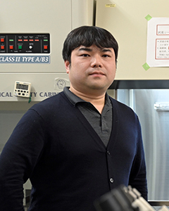
新生ポリペプチド鎖は、分子シャペロンや酸化異性化酵素など種々の因子により、正しい立体構造を獲得し、タンパク質としての機能を発揮する。しかし、細胞内では必ずしもタンパク質が正しい立体構造を獲得できる訳ではない。これら異常タンパク質の蓄積は、細胞内恒常性の破綻に繋がり、アルツハイマー病に代表される神経変性疾患や糖尿病、癌など様々な重篤な病態を惹起する。これら異常タンパク質に対するタンパク質品質管理・ストレス応答の分子機構を明らかにし、細胞内プロテオスタシスの解明を目指す。
主な業績
- Inoue, M., Sakuta, N., Watanabe, S., Zhang, Y., Yoshikaie, K., Tanaka, Y., Ushioda, R., Kato, Y., Takagi, J., Tsukazaki, T., Nagata, K., and Inaba, K. (2019) Structural Basis of Sarco/Endoplasmic Reticulum Ca 2+ -ATPase 2b Regulation via Transmembrane Helix Interplay. Cell Rep. 10.1016/j.celrep.2019.03.106
- Ushioda, R., and Nagata, K. (2019) Redox-mediated regulatory mechanisms of endoplasmic reticulum homeostasis. Cold Spring Harb. Perspect. Biol. 10.1101/cshperspect.a033910
- Maegawa, K., Watanabe, S., Noi, K., Okumura, M., Amagai, Y., Inoue, M., Ushioda, R., Nagata, K., Ogura, T., and Inaba, K. (2017) The Highly Dynamic Nature of ERdj5 Is Key to Efficient Elimination of Aberrant Protein Oligomers through ER-Associated Degradation. Structure. 25, 846-857.e4
- Ushioda, R., Miyamoto, A., Inoue, M., Watanabe, S., Okumura, M., Maegawa, K., Uegaki, K., Fujii, S., Fukuda, Y., Umitsu, M., Takagi, J., Inaba, K., Mikoshiba, K., and Nagata, K. (2016) Redox-assisted regulation of Ca2+ homeostasis in the endoplasmic reticulum by disulfide reductase ERdj5. Proc. Natl. Acad. Sci. 113, E6055–E6063
- Kawasaki, K., Ushioda, R., Ito, S., Ikeda, K., Masago, Y., and Nagata, K. (2015) Deletion of the collagen-specific molecular chaperone Hsp47 causes endoplasmic reticulum stress-mediated apoptosis of hepatic stellate cells. J. Biol. Chem. 290, 3639–46
- Ushioda, R., Hoseki, J., and Nagata, K. (2013) Glycosylation-independent ERAD pathway serves as a backup system under ER stress. Mol. Biol. Cell. 24, 3155–3163
- Ushioda, R., and Nagata, K. (2011) The Endoplasmic Reticulum-Associated Degradation and Disulfide Reductase ERdj5. Methods Enzymol. 490, 235–258
- Hagiwara, M., Maegawa, K., Suzuki, M., Ushioda, R., Araki, K., Matsumoto, Y., Hoseki, J., Nagata, K., and Inaba, K. (2011) Structural Basis of an ERAD Pathway Mediated by the ER-Resident Protein Disulfide Reductase ERdj5. Mol. Cell. 41, 432–444
- Ushioda, R., Hoseki, J., Araki, K., Jansen, G., Thomas, D. Y., and Nagata, K. (2008) ERdj5 is required as a disulfide reductase for degradation of misfolded proteins in the ER. Science. 321, 569–72
武田 洋幸(生命科学部 先端生命科学科 教授)
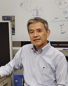
動物の発生は、発生ステージ特異的に特定の遺伝子群がゲノムより転写・翻訳されたタンパク質の複雑な相互作用によって制御されている。我々は、小型魚類(ゼブラフィッシュとメダカ)胚をモデルとし、発生遺伝学的・ゲノム科学的アプローチと最新のイメージング技術を駆使して、脊椎動物初期胚において体軸形成、器官形成に必須な分泌タンパク質の細胞外空間での拡散動態と活性制御機構をin vivo で明らかにする。また、脊椎動物のからだを構成する硬組織(骨格や耳石など)を生み出すバイオミネラリゼーションのメカニズムと進化の解明も目指す。
主な業績
- Fukushima HS, Ikeda T, Ikeda S, and Takeda H*. (2024)
Cell cycle length governs heterochromatin reprogramming during early development in non-mammalian vertebrates. EMBO Rep. 2024 Aug;25(8):3300-3323. (cover picture) - Fukushima HS, Takeda H*, and Nakamura R*. (2023)
Incomplete erasure of histone marks during epigenetic reprogramming in medaka early development. Genome Res. Apr;33(4):572-586. (cover picture) - Heilig AK, Nakamura R, Shimada A, Hashimoto Y, Nakamura Y, Wittbrodt J, and Takeda H*, Kawanishi T*. (2022)
Wnt11 acts on dermomyotome cells to guide epaxial myotome morphogenesis. Elife May 6;11:e71845. - Nakamura, R, Motai, Y, Kumagai, M, Wike, CL, Nishiyama, H, Nakatani, Y, Durand, NC, Kondo, K, Kondo, T, Tsukahara, T, Shimada, A, Cairns, BR, Aiden, EL, Morishita, S, and Takeda., H*. (2021).
CTCF looping is established during gastrulation in medaka embryos. Genome Res. 31(6):968-980. - Abe K. Shimada S. Tayama S, Nishikawa S, Kaneko T, Tsuda S, Karaiwa A, Matsui T, Ishitani T, and Takeda, H*/ Horizontal boundary cells, a special group of dermomyotomal cells, play crucial roles in the formation of dorsoventral compartments in teleost somite. (2019). Cell Rep. 27, 928–939.
- Yamaguchi H, Oda T, Kikkawa M*, and Takeda H*. (2018) Systematic studies of all PIH proteins in zebrafish reveal their distinct roles in axonemal dynein assembly. (2018) Elife May 9;7:e36979.
- Inoue Y, Saga T, Aikawa T, Kumagai M, Shimada A, Kawaguchi Y, Naruse K, Morishita S, Koga A, and Takeda., H.* (2017)
Complete fusion of a transposon and herpesvirus created the Teratorn mobile element in medaka fish. Nature Commun. 8, 551. - inoshita, M, Kamei, Y, Tamura, K, and Takeda, H*. (2013)
Trunk exoskeleton in teleosts is mesodermal in origin. Nat Commun. 4, 1639. - Omran H*, Kobayashi D, Olbrich H, Tsukahara T, Loges NT, Hagiwara H, Zhang Q, Leblond G, O'Toole E, Hara C, Mizuno H, Kawano H, Fliegauf M, Yagi T, Koshida S, Miyawaki A, Zentgraf H, Seithe H, Reinhardt R, Watanabe Y, Kamiya R, Mitchell DR*, and Takeda, H*. (2008)
Ktu/PF13 is required for cytoplasmic pre-assembly of axonemal dyneins. Nature 456, 611-6. - Horikawa, K*, Ishimatsu, K, Yoshimoto, E, Kondo, S, Takeda, H.* (2006)
Noise-resistant and synchronized oscillation of the segmentation clock. Nature 441, 719-23. (2006).


